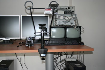
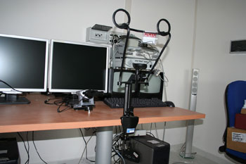
In the NNL Laboratory different instruments can be integrated (synchronized) to explore the neurophysiological activity of the brain during speech perception and production processing. Also, saccades and fixations of eye movements can be monitored during diversified audio-visual and reading tasks.
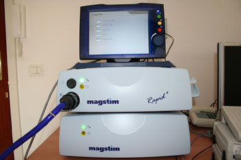
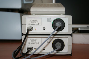
Repetitive Magnetic Transcranial Stimulation
(rMTS) with 64 channels: producing short magnetic impulses (of about 10 milliseconds) on the skull surface and particularly on the cerebral areas which are considered responsible for the tasks execution leads to interrupting (innocuously and temporarily) the functioning of the stimulated areas. Much better than PET and fMRI, rMTS allows the researcher to understand if the areas involved when accomplishing a cognitive activity are expressely destined to this activity.
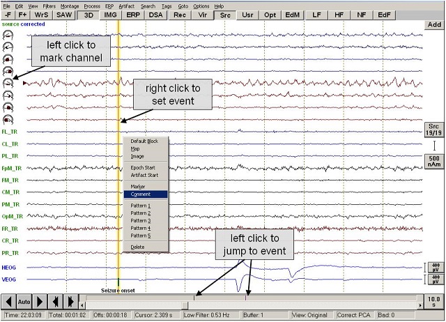
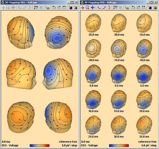
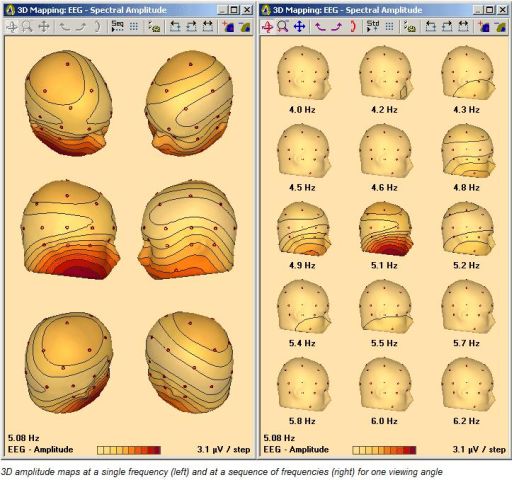
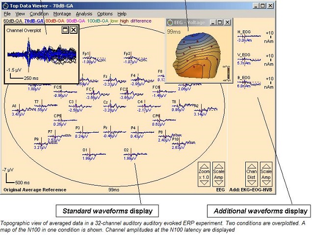
Event Related Potentials
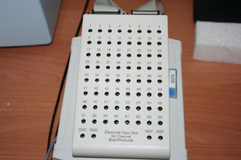
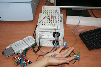
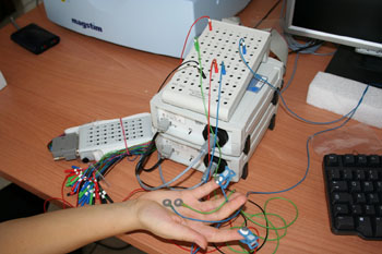
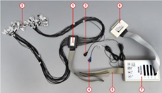
(ERPs) with 64 channels amplifier: it uses the classic electrophysiologic methods – based on the Electroencephalogram (EEG) – to produce small modifications in the spontaneous electric cerebral activity of a neural net. Such activity can be generated by means of an experimentally definable stimulous, i.e. a noun, a verb or a sentence, in order to investigate whether nouns and verbs produce different event-correlated potentials.
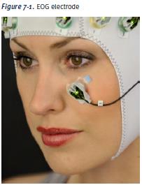
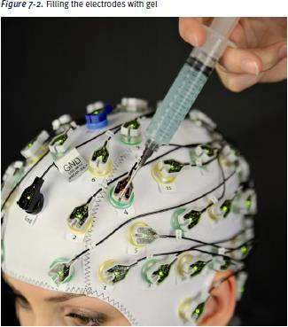
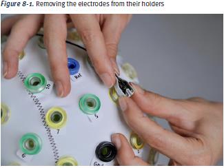
Event Related Potentials: actiCAP - The third generation of active electrodes
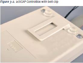
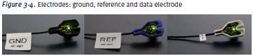
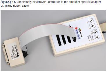
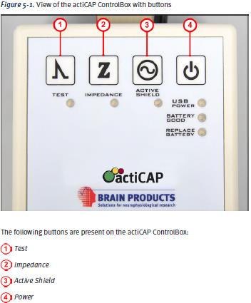
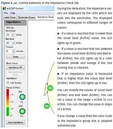
The actiCAP is a ground-breaking development aimed at raising EEG & ERP research to the next level. It combines active electrodes based on high-quality Ag/AgCl sensors with a new type of integrated noise subtraction circuits delivering even lower noise levels than any other “normal” active electrodes achieves. The actiCAP can be connected to nearly any existing research EEG amplifier system. When switching from passive to active electrodes, you will not need to exchange your entire recording system; simply keep the existing hard- and software, that you are used to. With the actiCAP you reduce your preparation time e.g. for 64 channels down to less than 10 minutes. Impedances are measured and displayed at each electrode by using LEDs Impedance values are stored in a text file and can be reviewed any time during data analysis Each electrode can be disconnected and replaced with a new one in case of malfunctioning Electrodes can be plugged into the cap before the cap is placed on the subject. In fact a slit in the electrode housing allows to inject gel and minimize the impedances while the cap and the electrodes are already in place. The actiCAP comes along with the latest version of actiCAP ControlSoftware
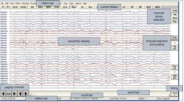
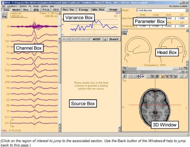
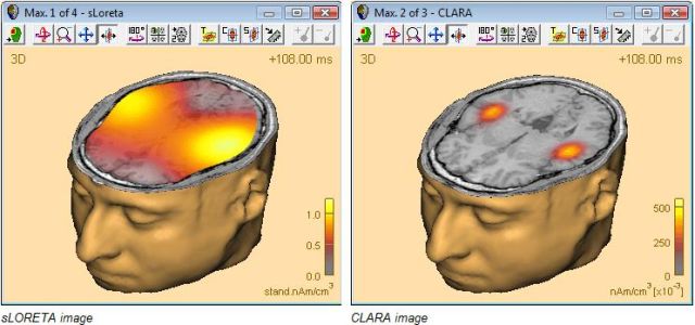
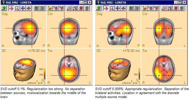
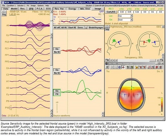
BESA Research Software
BESA Research is the most widely used software for source analysis and dipole localization in EEG, MEG, Evoked Potentials and ERP research. BESA Research has been developed on the basis of over 20 years experience in human brain research by Michael Scherg, University of Heidelberg, and Patrick Berg, University of Konstanz. BESA Research provides many advanced features for automatic source localization, fast modeling and easy, interactive hypothesis testing. Source analysis can be performed simultaneously on multiple conditions with advanced constraints based on anatomy and physiology. In addition to discrete multiple dipole modeling, all major distributed imaging methods are also available for comparison. This makes BESA a complete all-in-one tool for source imaging. BESA has all the features required to perform offline processing of continuously acquired EEG and MEG data using external and internal triggers, e.g. generated from an EMG channel or by spatio-temporal pattern search. Triggers are automatically mapped into predefined paradigms to provide for fast selection of combinations of averages (addition, subtraction, subsets). Based on the spatial components approach of Berg and Scherg (1994), artifacts (e.g. eye and ECG) can be corrected on the fly from EEG, MEG, and ERP data. Special viewing options allow for easy selection of averaged or single epochs of interest (e.g. spikes) and for immediate source localization and analysis. Digitized 3D sensor locations can be used to coregister EEG or MEG data with structural and functional MRI. Fitted dipole sources can be superimposed directly to the individual MR image.
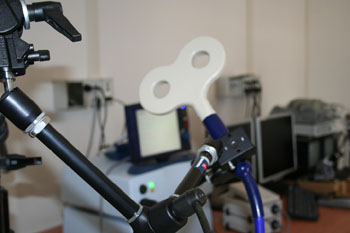
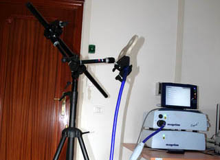
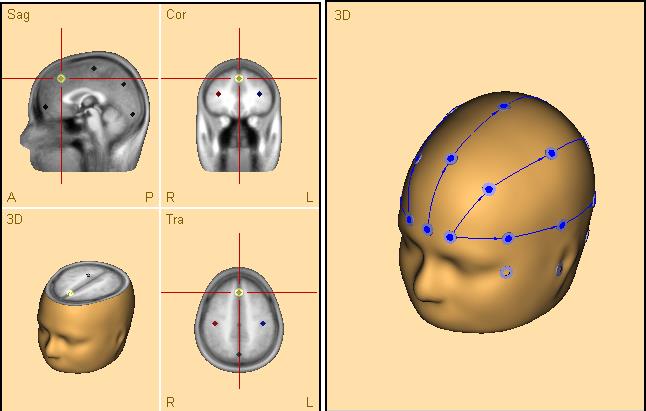
Softaxic System
it allows the researcher to place the coil of the magnetic stimulator on the cerebral cortex of the speaker, with or without magnetic resonance. It offers a 3D reconstruction and the stereotaxic navigation in human brain.
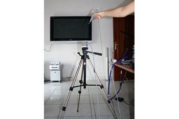
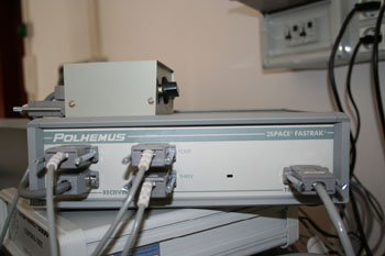
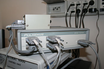
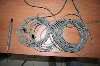
Spatial Digitalization System
it allows the researcher to monitor the movements of the head, of the hands as well as to monitor tools suited to biomechanics analysis, to stereotaxic localization, to telerobotics and to virtual reality applications.
Presentation Software
it enables the researcher to create personalized auditory and visual stimuli.
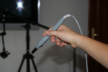
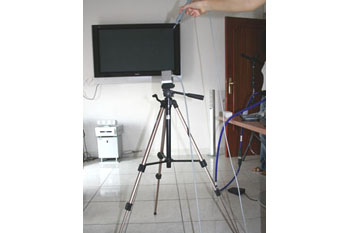
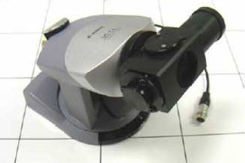
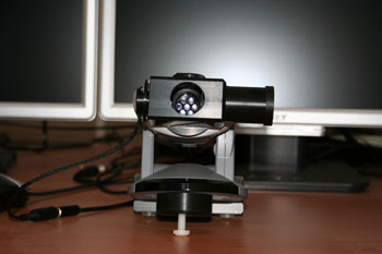
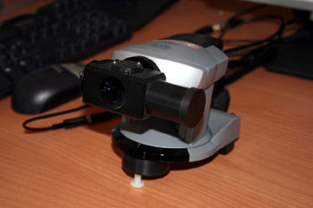
Eye Tracker
it is an infrared-rays system suited to studying the eye and saccadic movements and equipped with remote optics and monitoring system of framing and inclination (working at 120, 240, e 360Hz). It allows the informant to move his head freely in 30 square centimetres and it does not need to dispose of a support to block the informant head.